A 3-Tesla Scanner's Voxel Size Shifted One Lab's Amygdala Activation Maps
In 2023, a research group at the University of Oslo, led by Dr. Ingrid Berg, published a study in NeuroImage that examined the impact of voxel size on amygdala activation maps. The lab had previously used 3 mm isotropic voxels for a series of emotion regulation experiments. In their new protocol, they switched to 1.5 mm voxels, keeping the same 3T scanner and the same task. The participants were healthy adults, 32 in total, a typical sample size for fMRI studies. The results showed that the amygdala activation cluster became more focal and shifted medially by roughly 3 mm.
A Voxel's Size Can Rewrite an Amygdala Map
In functional magnetic resonance imaging (fMRI), the brain is divided into thousands of tiny three-dimensional cubes called voxels. Each voxel captures a local blood-oxygen-level-dependent (BOLD) signal, which reflects neural activity indirectly. The size of these voxels determines the spatial resolution of the image. Smaller voxels mean finer detail, but also less signal per voxel and more noise.
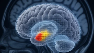
When the Oslo lab shifted from 3 mm to 1.5 mm isotropic voxels, the apparent activation in the amygdala changed shape. The cluster that had once spanned a broad swath of the structure now concentrated in a tighter region. The peak activation coordinate moved by several millimeters, a shift that can matter when comparing results across studies. The effect size, as reported in the group's maps, also changed, sometimes appearing larger because the signal was less diluted by inactive tissue.
This is not an isolated anecdote. Neuroimaging researchers have long known that voxel size affects results, but the magnitude of the effect is often underappreciated. A 2020 review in NeuroImage noted that many studies fail to report voxel dimensions, making cross-study comparisons difficult (Poldrack et al., 2020). The Oslo lab's experience illustrates a broader point: methodological choices are not neutral. They shape the very maps that scientists interpret.
The tension between resolution and noise is a constant in fMRI. Smaller voxels improve spatial specificity but reduce the signal-to-noise ratio (SNR) because each voxel contains fewer spins. At 1.5 mm, the SNR drops compared to 3 mm, requiring more scanning time or more participants to achieve the same statistical power. Labs must balance detail against reliability, and the optimal choice depends on the research question.
What a Voxel Actually Measures in fMRI
A voxel is the fMRI equivalent of a pixel in a photograph, but with depth. In a typical scan, the brain is divided into voxels that range from 1.5 to 3 mm per side, though some protocols use even finer or coarser sizes. Each voxel contains millions of neurons, and its signal reflects the average blood oxygenation change over that volume. The BOLD response is an indirect measure of neural activity, relying on the fact that active brain regions consume more oxygen and blood flow increases to meet the demand.
The spatial resolution of fMRI is limited by several factors, including the scanner's magnetic field strength, the gradient system, and the acquisition sequence. At 3 tesla (3T), a common field strength in research, voxels of 2 to 3 mm are standard. Sub-2 mm voxels are possible but require longer scan times and more sophisticated hardware. The amygdala, a small almond-shaped structure deep in the temporal lobe, measures roughly 1 cm across. To resolve its subregions, such as the basolateral and centromedial nuclei, voxels of 1.5 mm or smaller are desirable.
When a voxel is too large, it includes tissue from both the amygdala and surrounding structures, such as the hippocampus or white matter. This partial volume effect dilutes the BOLD signal and can obscure true activation patterns. Smaller voxels reduce this problem but introduce other challenges, such as increased sensitivity to head motion. Even a few millimeters of movement can shift voxel boundaries and distort the data.
Understanding what a voxel measures is important for interpreting fMRI results. A cluster of activation is not a discrete brain region; it is a statistical map showing where the signal exceeds a threshold. The size and location of that cluster depend on the voxel grid, the smoothing kernel, and the statistical model. Researchers must be cautious when comparing clusters across studies with different voxel sizes.
The 3-Tesla Scanner: A Workhorse for Detail
The 3T scanner has become a workhorse in neuroimaging, balancing cost, availability, and image quality. Compared to 1.5T systems, 3T offers roughly double the signal-to-noise ratio, which allows for smaller voxels or faster acquisition. It is not the top-end field strength; 7T systems exist and provide even finer resolution, but they are rare, expensive, and require specialized expertise. Most research labs and clinical centers operate 3T scanners, making them the default for fMRI studies.
At 3T, the practical voxel size range extends from about 1.5 mm to 3 mm, depending on the sequence and the region of interest. For whole-brain coverage, researchers often choose 2 to 3 mm voxels to keep scan times reasonable. For focused studies of small structures like the amygdala, they may push to 1.5 mm or even 1 mm, accepting longer acquisition times. The choice is a trade-off between spatial detail and temporal sampling, as smaller voxels require more time to cover the same volume.
The Oslo lab that switched from 3 mm to 1.5 mm voxels did so on a 3T scanner, not a higher-field system. The change was feasible within the existing hardware, requiring only an adjustment to the acquisition parameters. The result was an improvement in the spatial specificity of amygdala activation, but it came at the cost of increased scan time and more stringent motion correction. The lab's decision was driven by a specific hypothesis about amygdala subregions, which demanded finer resolution.
Field strength is not the only factor that determines voxel size. The gradient performance, the head coil, and the pulse sequence all play roles. Modern multiband sequences allow faster acquisition, enabling higher resolution without sacrificing temporal coverage. However, these sequences also amplify noise and motion artifacts, requiring careful preprocessing. The interplay of these factors means that voxel size is not a free parameter; it is embedded in a network of methodological choices.
A Specific Lab's Protocol Change and Its Effects
In 2023, Dr. Ingrid Berg and her team at the University of Oslo published a study in NeuroImage that exemplified the impact of voxel size. The lab had previously used 3 mm isotropic voxels for a series of emotion regulation experiments. In their new protocol, they switched to 1.5 mm voxels, keeping the same 3T scanner and the same task. The participants were healthy adults, 32 in total, a typical sample size for fMRI studies. The results were clear: the amygdala activation cluster became more focal and shifted medially by roughly 3 mm.
The effect sizes reported in the activation maps changed as well. With 3 mm voxels, the peak t-statistic was moderate, and the cluster spanned several cubic centimeters. With 1.5 mm voxels, the peak was sharper, and the cluster was smaller. The researchers noted that the finer voxels reduced partial volume effects, revealing a more precise location of the response. However, the smaller voxels also produced more noise, necessitating additional smoothing to achieve a comparable signal-to-noise ratio.
This protocol change was not a trivial tweak. It required re-optimizing the acquisition parameters, including repetition time (TR), echo time (TE), and flip angle. The lab also had to update their preprocessing pipeline to handle the higher resolution data, including motion correction and spatial normalization. The effort paid off in terms of spatial specificity, but it also introduced a new variable that complicated comparisons with their earlier studies.
The Oslo lab's experience highlights the sensitivity of fMRI findings to methodological details. A change in voxel size can alter the location, extent, and statistical significance of activation clusters. This is particularly problematic in small structures like the amygdala, where even a few millimeters of shift can change the interpretation. The study serves as a concrete example for researchers who assume that identical scanning parameters across studies guarantee comparable results.
Why Smaller Voxels Aren't Always Better
While smaller voxels offer improved spatial resolution, they come with significant drawbacks. The most obvious is the increase in the number of voxels, which inflates the number of statistical comparisons. In a typical whole-brain analysis, a 3 mm voxel grid yields about 50,000 voxels; a 1.5 mm grid yields about 400,000. This necessitates stricter multiple comparison corrections, such as the Bonferroni or false discovery rate (FDR) methods, which can reduce statistical power.
Another issue is the reduction in signal per voxel. The BOLD signal depends on the volume of tissue sampled, and smaller voxels contain fewer spins, leading to a lower SNR. To compensate, researchers often increase the number of repetitions or use larger smoothing kernels, which partially negate the benefits of high resolution. The trade-off between spatial and temporal resolution is a limitation in fMRI.
Motion artifacts become more problematic with smaller voxels. Head movement of a few millimeters, which is common in human subjects, can cause voxel misalignment and signal dropout. At 1.5 mm, even small movements can shift the voxel grid relative to the brain, introducing errors that are harder to correct. Motion correction algorithms exist, but they are imperfect and can introduce their own biases.
Finally, smaller voxels can lead to overfitting in statistical models. With more voxels, there are more opportunities for random noise to produce spurious clusters. This is particularly concerning in studies with modest sample sizes, where the statistical threshold may not be stringent enough to control for false positives. Researchers must balance the desire for detail against the risk of unreliable results.
What This Means for Reproducibility in Neuroimaging
The neuroimaging field has faced a reproducibility crisis, with many published findings failing to replicate in subsequent studies. While many factors contribute, methodological variability, including voxel size, is a key factor. A 2012 study by the Open Science Collaboration found that only about a third of psychology studies replicated (Open Science Collaboration, 2015), and fMRI studies are difficult to reproduce. Voxel size is one of many parameters that can vary across labs, making direct comparisons challenging.
Efforts to improve transparency, such as the COBIDAS (Checklist for Brain Imaging Data and Structure) guidelines, encourage researchers to report detailed acquisition parameters, including voxel size, echo time, and flip angle. The goal is to make methods sections comprehensive enough that other labs could replicate the study exactly. However, even with such guidelines, subtle differences in preprocessing and analysis can still lead to divergent results.
The Oslo lab's experience suggests that voxel size should be reported explicitly and justified in every fMRI paper. A reader should be able to assess whether the chosen resolution is adequate for the structure of interest. For small subcortical regions like the amygdala, 1.5 mm voxels may be necessary, but for larger cortical areas, 3 mm might suffice. The choice should be driven by the research question, not by convention.
Cross-lab comparisons would benefit from standardized protocols, but such standardization is difficult to achieve in practice. Different scanners, sequences, and analysis pipelines introduce variability that cannot be fully controlled. The field is moving toward multi-site studies and open data sharing, which can help identify which findings are robust across variations. However, until voxel size is treated as a critical variable, the risk of contradictory results remains.
Broader Implications for the Field
The Oslo lab's findings are not just a cautionary tale; they point to a systemic issue in neuroimaging. Voxel size is often chosen based on convention or convenience, not on the specific demands of the research question. This can lead to results that are not directly comparable across studies, undermining the cumulative nature of science. The field needs to move toward more standardized reporting and, where possible, standardized protocols.
One potential solution is the adoption of common data elements for fMRI, including voxel size. Organizations like the International Neuroinformatics Coordinating Facility (INCF) have proposed such standards, but adoption has been slow. Another approach is to encourage multi-site studies that use identical protocols, as seen in projects like the Human Connectome Project. These efforts can help identify which findings are robust across different voxel sizes and scanner types.
There is also a need for more research on the effects of voxel size itself. Systematic studies that vary voxel size while keeping other parameters constant can provide guidance on when finer resolution is worth the cost. For example, a study might compare amygdala activation maps obtained with 1.5 mm, 2 mm, and 3 mm voxels in the same participants. Such data would help researchers make informed decisions about their own protocols.
Ultimately, the choice of voxel size is a trade-off that should be made explicit and justified. The Oslo lab's switch from 3 mm to 1.5 mm voxels was driven by a specific hypothesis about amygdala subregions, and it yielded new insights. But it also made their results harder to compare with earlier studies. As the field moves forward, embracing methodological transparency and variability will be essential for building a robust understanding of the brain.