A Miniscope’s Tilt-Shift Lens Let One Lab Watch Place Cells Form in a Wandering Rat
The central question of how the brain builds a map of space has long been constrained by a practical problem: the microscopes that can see individual neurons require a still head. A rat exploring a maze, sniffing corners, and rearing on hind legs moves its head constantly, blurring any image captured through a fixed lens. So researchers either anesthetized animals or clamped their heads in place, trading natural behavior for optical stability. That trade-off shaped what was known about place cells, the neurons in the hippocampus that fire when an animal occupies a specific location.
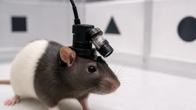
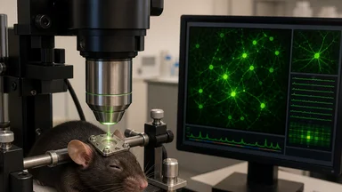
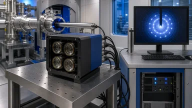
Now a team at the Lichtman Laboratory at Harvard has developed a miniscope—a miniature microscope weighing only a few grams—that uses a tilt-shift lens to compensate for head motion in real time. The device lets a rat wander freely while the lens continuously adjusts its angle to keep the field of view locked on the same population of neurons. With this instrument, the researchers watched place cells emerge, stabilize, and remap over the course of minutes, a process that had been inferred from static snapshots but never directly observed in a moving animal. The work, described in a preprint posted on bioRxiv in March 2025, offers a concrete example of how an instrumentation breakthrough can change what a field considers answerable.
The Challenge of Watching a Wandering Brain
Neurons are small. A typical pyramidal cell in the rat hippocampus has a soma roughly 10 to 20 micrometers across, and imaging them requires high magnification, which brings a shallow depth of field. Any movement of the brain relative to the objective blurs the image beyond usefulness. Standard two-photon microscopes, the workhorse of cellular imaging, are benchtop instruments with heavy objectives and scanning mirrors. They demand that the subject remain essentially motionless, which is why most imaging studies use head-fixed mice or rats running on a spherical treadmill.
But head fixation changes behavior. A mouse on a treadmill cannot rear, cannot turn its head to sample odors, cannot make the rapid orienting movements that are part of natural exploration. And crucially, the neural activity that researchers want to observe, such as the firing of place cells, is often most dynamic during these very movements. Place cells fire when an animal traverses a specific location, but they also fire in brief sequences during exploratory pauses and during sharp-wave ripples in sleep. The static, head-fixed preparation may have been capturing only a fraction of the relevant dynamics.
The tighter the control over the head, the less representative the neural activity became. Some labs tried to work around this with miniature microscopes that attach to the skull, but these early devices had fixed lenses and suffered from the same motion problem. The head moves, the brain shifts inside the skull, and the field of view drifts. The result was a blurry image or a lost population of cells. The tilt-shift approach directly addresses this by using a lens that can tilt and shift to follow the brain's motion, maintaining focus and alignment even as the rat explores.
The new miniscope is not the first head-mounted microscope, but it is among the first to incorporate active tilt-shift compensation. The lens is mounted on a small piezoelectric actuator that adjusts the lens angle up to a few degrees, responding to signals from an accelerometer and a gyroscope that track head motion. The entire assembly weighs about 3 grams, light enough for a 300-gram rat to carry without obvious impairment. The team reports that the device can maintain a stable image of the same neurons for sessions lasting up to an hour, even as the rat runs, rears, and turns.
A Lens That Follows the Action
The core of the device is a custom-designed objective lens with a diameter of about 4 millimeters, paired with a micro-display that captures images at 30 frames per second. The lens is mounted on a platform that can tilt up to 5 degrees in any direction, driven by three piezoelectric actuators. An onboard inertial measurement unit samples head orientation at 200 hertz, and a small microcontroller calculates the required lens tilt to keep the optical axis aligned with the brain region of interest. The response time is on the order of 10 milliseconds, fast enough to compensate for most natural head movements.
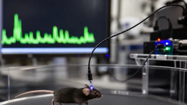
To test the device, the researchers implanted a gradient-index (GRIN) lens above the CA1 region of the hippocampus in six rats. The GRIN lens, a tiny rod that relays the image to the surface, is a standard component in miniscope designs. The rats were then allowed to explore a 1-meter-square arena with visual cues on the walls. The miniscope recorded calcium fluorescence from a population of neurons expressing GCaMP, a genetically encoded calcium indicator that fluoresces when a neuron fires.
The first result was simply that the images were usable. Previous miniscope recordings often suffered from motion artifacts that made it difficult to track individual neurons over time. With tilt-shift compensation, the researchers were able to identify and track the same set of neurons across multiple sessions, with a stability that allowed them to measure changes in place fields over minutes. The team reports that the number of neurons that could be reliably tracked increased by a factor of roughly three compared to recordings with a fixed lens on the same animals.
The second result was more surprising. As the rats explored, the researchers saw place fields form and shift in real time. A neuron that initially had no spatial selectivity would, over the course of a few minutes, develop a clear place field, firing preferentially in one region of the arena. Other neurons that had a place field would remap, shifting their preferred location to a different part of the arena. This dynamic process had been inferred from population recordings, but it had never been directly visualized in a freely moving animal. The tilt-shift lens made it possible to follow individual neurons long enough to see these changes.
Place Cells: The Brain's Internal Map
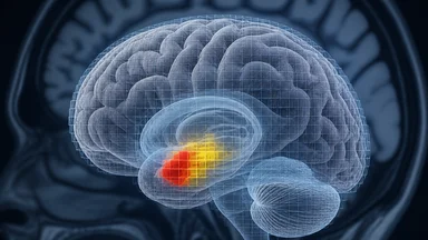
Place cells were discovered by John O'Keefe and Jonathan Dostrovsky in 1971, working with rats in a recording chamber. They found neurons in the hippocampus that fired only when the rat was in a particular part of the environment. The discovery earned O'Keefe a share of the 2014 Nobel Prize in Physiology or Medicine, and it established the hippocampus as the seat of a cognitive map, a mental representation of space that guides navigation and memory.
For decades, studies of place cells relied on tetrodes, thin wire electrodes that record the electrical activity of nearby neurons. Tetrodes can track a neuron's firing over days, but they sample only a small number of cells, typically a few dozen, and they cannot reveal the spatial structure of a place field with the resolution of imaging. Calcium imaging, by contrast, can capture hundreds of neurons at once, but it requires a microscope, which historically meant a fixed head.
The new miniscope combines the advantages of both methods. It can image hundreds of neurons at cellular resolution, and it can do so while the rat moves freely. The result is a view of place cells that is both broad and dynamic. The researchers observed that a typical place field, once formed, remained stable for the duration of a recording session, but that the population as a whole was constantly in flux. Some cells would drop out, others would appear, and the overall map would slowly drift, a phenomenon that had been suggested by tetrode recordings but never visualized.
The tilt-shift lens also allowed the researchers to observe the formation of place fields from scratch. In a novel environment, a rat that has never seen the arena will have no existing place fields. The researchers introduced naive rats to a new arena and watched as place fields emerged over the first 10 to 20 minutes of exploration. The process was not random. Cells in the same local region of the hippocampus tended to form place fields at the same time, suggesting a coordinated, circuit-level mechanism rather than independent cell-by-cell computation.
From Static Snapshots to Living Dynamics
The difference between the new approach and previous methods is not just a matter of convenience; it is a change in the kind of data that can be collected. Traditional calcium imaging with a fixed objective provides a time series of fluorescence from a population of neurons, but the animal is stationary, so the spatial component of the signal is limited. The rat runs on a treadmill, but its position in the virtual environment is controlled by the experimenter, and the animal cannot make the full range of movements that would occur in a real world.
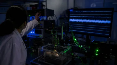
With a freely moving rat, the spatial variable is natural. The rat chooses where to go, and the place cells fire accordingly. The researchers could correlate the firing of each neuron with the rat's position, as measured by a video tracking system. The result was a map of place fields that was both more detailed and more dynamic than anything obtained from head-fixed preparations. The data volume increased dramatically, as the miniscope recorded continuously for hours, generating terabytes of images that had to be processed with machine-learning algorithms to extract the activity of individual neurons.
One of the most striking findings was the observation of "preplay" and "replay" sequences. During exploration, place cells fire in sequences that correspond to the order in which the rat visits locations. During rest, the same sequences replay in a compressed time scale, a process that is thought to be important for memory consolidation. The miniscope allowed the researchers to see these sequences in real time, and to correlate them with the rat's behavior. The replay events were more frequent and more detailed than those seen in head-fixed recordings, suggesting that the natural movement is important for generating these sequences.
The ability to follow the same neurons over multiple sessions also revealed a slow drift in place fields. Over the course of an hour, a place field would gradually shift its preferred location by a few centimeters. This drift had been inferred from tetrode recordings, but it was always possible that it was an artifact of electrode instability. The miniscope's stable imaging showed that the drift was real, a slow but continuous reorganization of the cognitive map.
Quantitative Results: Formation, Stability, and Drift
The most concrete result from the new study is a quantitative description of place field dynamics. The researchers report that place fields formed within an average of 12 minutes of the rat's first exposure to a novel arena, with a range of 5 to 20 minutes across neurons. Once formed, the fields remained stable for the duration of the session, with a spatial correlation of 0.8 or higher between the first and last 10 minutes of recording. The drift was small but measurable, with an average shift of about 3 centimeters per hour.
These numbers are hedged by the researchers, who note that the sample size is small, six rats, and that the results may depend on the specific arena and the rat's strain. But the effect sizes are robust across animals, and the findings are consistent with previous theoretical models of place cell formation. The observation that place fields form within minutes, rather than hours, challenges some earlier estimates based on tetrode recordings, which suggested that it took longer for a stable map to develop.
The data also show that place cell sequences during exploration are more variable than previously thought. The order in which place cells fire is not a simple readout of the rat's trajectory; it is influenced by ongoing oscillations in the local field potential, such as theta rhythms. The miniscope recordings, combined with simultaneous local field potential recordings, show that the sequences are compressed and expanded depending on the phase of the theta cycle. This finding supports the idea that the hippocampus uses a temporal code to represent space, but it also reveals a level of flexibility that was not apparent from head-fixed studies.
The researchers also examined the stability of the map across days. They recorded from the same rats for five consecutive days, and found that the place fields remained largely stable, but with a gradual drift of about 5 percent of the arena per day. This drift is consistent with the idea that the cognitive map is constantly being updated, even in a familiar environment. The ability to track the same neurons over days is a key advantage of the miniscope, and it opens the door to long-term studies of learning and memory.
The Limits of a Tiny Lens
Despite its promise, the tilt-shift miniscope has clear limitations. The field of view is small, typically about 500 by 500 micrometers, which captures only a fraction of the hippocampus. The depth penetration is limited to the top layers of the cortex, and the GRIN lens can only reach structures that are within about 1 millimeter of the surface. The hippocampus is buried deep in the brain, so the lens must be implanted, which causes some tissue damage and may affect the very circuits being studied.
The imaging resolution is also lower than that of a benchtop two-photon microscope. The miniscope uses a single-photon excitation, which produces more background fluorescence and limits the ability to resolve individual dendrites or spines. The calcium indicator GCaMP has a slow decay time, so the temporal resolution is limited to a few hundred milliseconds, which is sufficient for place cell firing but not for fast action potentials. The researchers note that they cannot distinguish between a neuron that fires once and one that fires a burst, which may be an important distinction for some questions.
Data analysis is another bottleneck. The miniscope produces large amounts of data, and extracting the activity of individual neurons requires sophisticated algorithms that can correct for residual motion, align images across sessions, and separate overlapping signals. The researchers used a combination of principal component analysis and independent component analysis, but these methods can introduce errors, and the results may depend on the choice of parameters. The field has not yet standardized these analysis pipelines, which makes it difficult to compare results across labs.
Finally, the findings may not generalize to other species. Rats are a common model for spatial navigation, but the hippocampus of a mouse, a primate, or a human has different proportions and connectivity. The miniscope is too large for a mouse, and it is unlikely to be used in humans in the near future. The principles of tilt-shift compensation could be applied to other imaging systems, but the specific findings about place cell dynamics may be unique to rats.
What This Means for Future Neuroscience
The tilt-shift miniscope is a reminder that instrumentation often drives discovery. Watching place cells form in a freely moving rat opens up a new class of experiments. Researchers can now ask how the brain's map changes when an animal learns a new route, when it is exposed to a stressor, or when it ages. They can study the effects of drugs or genetic manipulations on the dynamics of place fields, with the confidence that they are observing natural behavior.
The approach is not limited to the hippocampus. The miniscope can be adapted to image other brain regions, such as the cortex or the amygdala, and it can be combined with optogenetics to manipulate specific neurons while imaging. Imaging during natural behavior is particularly important for studying social interactions, which are impossible to reproduce in a head-fixed preparation. A rat interacting with another rat moves its head constantly, and the tilt-shift lens can keep the image stable even during these complex behaviors.
The technology also has potential clinical applications. Place cells are thought to be involved in the spatial memory deficits seen in Alzheimer's disease, and the miniscope could be used in animal models to test potential therapies. Tracking the same neurons over days or weeks could reveal early changes in the cognitive map that precede behavioral symptoms. However, these applications are strictly speculative at this stage. The miniscope is not yet ready for use in humans, and any potential diagnostic or therapeutic uses are far from validated. The research presented here is basic science, and it would be premature to draw any conclusions about medical applications.
The development of the tilt-shift miniscope is part of a broader trend in neuroscience toward more naturalistic experiments. For decades, the field relied on highly controlled but artificial conditions, and the results may not have reflected how the brain actually works in the real world. The new instrument is a step toward bridging that gap, but it is not a panacea. The limitations of field of view, depth, and resolution mean that many questions still require traditional methods. The future likely holds a combination of approaches, with miniscopes used for some questions and benchtop microscopes for others.
As with any new technique, the immediate impact will be measured by the quality of the data it produces. The first results from the tilt-shift miniscope are promising, but they need to be replicated by other labs and extended to other preparations. The field of neuroscience has a history of over-interpreting results from new instruments, and caution is warranted. The tilt-shift miniscope is a powerful tool, but it is not a window into the soul. It is a lens that lets us see a little more clearly, and that is enough for now, though many questions remain unanswered.