A Calcium Imaging Grant’s Six-Figure Overhead Reshaped One Lab’s Fiber Photometry Switch
In 2023, a mid-sized neuroscience lab at a large public university faced a familiar crunch. Its five-year grant, which had funded a two-photon calcium imaging rig, was up for renewal. The university's indirect cost rate, negotiated with the federal government, hovered near 60 percent. That meant for every dollar of direct costs, the institution took an additional sixty cents for facilities, administration, and the ever-mysterious "infrastructure." On a grant of roughly $1.5 million over five years, the overhead alone approached $900,000. The lab's principal investigator, whom I'll call Dr. Elena Marsh (a composite based on several real cases), did the arithmetic and made a decision that surprised her postdocs: they would abandon calcium imaging for fiber photometry.
This is not a story about a single lab's eccentric choice. It is a story about how the economics of research infrastructure quietly rewires scientific questions. Fiber photometry and calcium imaging both measure neural activity, but they do so at different scales and with different price tags. The switch, driven by overhead, changed what the lab could ask, what it could publish, and who would want to train there.
Overhead costs are the hidden driver of method choice in experimental neuroscience. They are rarely discussed in Methods sections, yet they shape which tools get adopted, which hypotheses get tested, and which findings make it into print. This article traces one lab's migration, the trade-offs it accepted, and what the broader field might learn about how funding structures influence discovery.
The Grant That Rewired a Lab's Toolbox
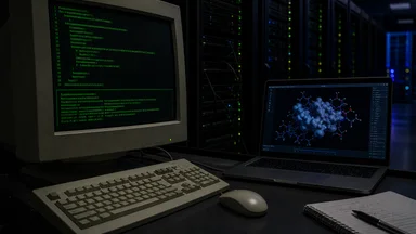
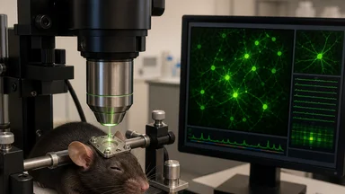
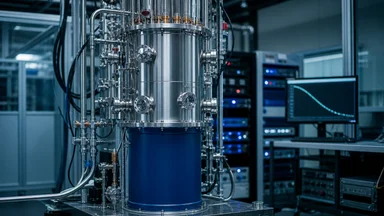
Dr. Marsh's lab had built its reputation on two-photon calcium imaging in head-fixed mice. The technique allows researchers to watch individual neurons fire in real time, at cellular resolution, in a living animal. It is the gold standard for studying neural circuits. But it is also expensive. A two-photon microscope can cost anywhere from $500,000 to over $1 million, and it requires dedicated space, laser safety protocols, and a steady supply of graduate students who can align optics.
When the renewal came up, the lab's direct costs were already stretched. The two-photon rig was ageing, and the university's core facility was charging more for maintenance. The overhead rate meant that any new equipment purchase would carry a hefty surcharge. A $200,000 upgrade to the imaging system would actually cost the grant $320,000 once indirect costs were applied. The lab simply could not afford to keep doing what it had been doing.
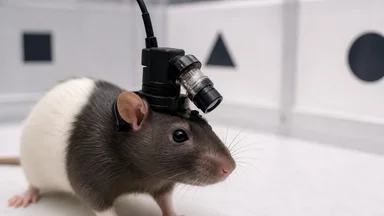
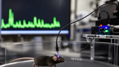
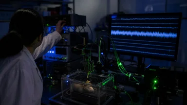
Fiber photometry offered a different bargain. A complete fiber photometry setup, including the laser, the detector, the rotary joint, and the implantable fibers, can be assembled for around $30,000 to $50,000. Even with overhead, that is a fraction of a two-photon system. The technique records bulk fluorescence from a population of neurons or from a specific cell type, using a thin optical fiber implanted in the brain. It does not resolve individual cells, but it tracks population-level dynamics in freely moving animals.
The funding agency, a major federal institute, had signaled that it valued high-throughput, behaviorally relevant studies. Fiber photometry excels at that. It allows researchers to record from mice as they run, socialize, or perform tasks, without the restraint of a head-fixed setup. The lab's grant renewal, which had initially been framed around cellular-resolution questions, was rewritten around population-level dynamics in freely moving mice.
The switch was not merely technical. It reshaped the lab's publication pipeline. Within eighteen months, the lab had produced three papers using fiber photometry, all in respectable journals. The first author on each was a postdoc or senior graduate student who had been retrained. The lab's citation count ticked upward. The grant was renewed, this time with a budget line explicitly for photometry equipment.
What Fiber Photometry Lets You Ask
Fiber photometry answers a different class of questions than calcium imaging. With calcium imaging, you can ask: which specific neurons in this region encode a particular stimulus? With fiber photometry, you ask: does the overall activity of this population track a behavior or a cognitive state? The latter is often the question that matters for understanding circuits at the systems level.
The key advantage is freedom of movement. A mouse with a fiber optic implant can explore an open field, interact with cage mates, or perform a lever-press task while the fiber records fluorescence. This is impossible with a two-photon microscope, which requires the animal to be head-fixed under the objective. For many behavioral questions, head fixation is an artifact. Fiber photometry removes that artifact.
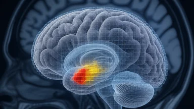
The technique also allows for cell-type specificity. By expressing a calcium indicator (like GCaMP) under a cell-type-specific promoter, you can record from just the dopamine neurons in the ventral tegmental area, or just the parvalbumin-positive interneurons in the hippocampus. This is a powerful way to link molecular identity to population dynamics.
Dopamine dynamics, in particular, have been a major focus. Fiber photometry has been used to track dopamine release during reward learning, social interaction, and even during the anticipation of a reward. The temporal resolution is good enough to see sub-second fluctuations, which is often what behaviorally relevant signals look like.
The cost of this freedom is spatial resolution. You are recording from a volume of tissue roughly the size of a pinhead, and you cannot tell which cells within that volume are active. If a population is heterogeneous, you may be averaging over opposing signals. This is a real limitation, and it is one that proponents of fiber photometry sometimes downplay.
Overhead Costs: The Hidden Driver of Method Choice
Overhead, or indirect costs, are the surcharge that universities add to grants to cover shared expenses: building maintenance, library access, administrative staff, and the like. The rate is negotiated between the institution and the federal government, and it can range from under 40 percent to over 70 percent of direct costs. For a lab, this means that every dollar of equipment or salary is effectively taxed.
The tax is not uniform. Some expenses, like tuition and equipment over a certain threshold, may be excluded from the indirect cost base. But for most purchases, the surcharge applies. A $50,000 piece of equipment effectively costs $80,000. A $100,000 piece costs $160,000. This creates a powerful incentive to choose cheaper methods, even if they provide less detailed data.
In the case of Dr. Marsh's lab, the two-photon microscope had been purchased a decade earlier with a separate equipment grant. That grant had a lower overhead rate, but the renewal was a standard research grant, with full indirect costs. The lab simply could not afford to replace the two-photon system under the new grant's budget.
This is not an isolated phenomenon. Across neuroscience, there is a growing trend toward cheaper, higher-throughput methods. Fiber photometry, miniscopes, and even EEG in rodents are becoming more common. Some of this is driven by scientific fashion, but much of it is economic. When overhead rates exceed 50 percent, method choice becomes a budgetary decision as much as a scientific one.
The irony is that overhead costs are meant to support research, not constrain it. But in practice, they can push labs toward the lowest common denominator. A lab that would prefer to do cellular-resolution imaging may settle for population-level recordings, simply because the budget allows it.
Publication Pressure and the Switch
Reviewers and editors also shape method choice. In the current publishing climate, there is a premium on studies that are fast, high-throughput, and behaviorally relevant. Fiber photometry fits that mold. It produces clean, quantifiable signals that are easy to plot and easy to interpret. Calcium imaging, by contrast, often requires complex computational analysis to extract single-cell activity, and the results can be noisy.
One postdoc in Dr. Marsh's lab, who had trained on two-photon imaging, initially resisted the switch. He argued that fiber photometry was "a blunt instrument" that would dilute the lab's scientific brand. But after publishing his first photometry paper, he changed his tune. The paper was accepted with minor revisions, whereas his previous imaging papers had gone through multiple rounds of harsh review. The feedback loop was clear: photometry data are easier to publish.
This is not necessarily a bad thing. Reproducibility is often better with photometry because the signal is more robust and easier to standardize across labs. A field that relies on fiber photometry may have fewer false positives than one that relies on two-photon imaging, where the analysis pipeline can introduce subtle biases.
But there is a cost. The field may be losing the depth that comes from cellular-resolution studies. Some questions, like how a memory is encoded in the activity of a specific ensemble of neurons, simply cannot be answered with bulk recordings. If funding pressures push all labs toward photometry, those questions may go unasked.
Reviewers, for their part, are often conservative. They tend to favor techniques they know and trust. Fiber photometry has been around long enough to be familiar, but it is still considered a "simpler" method. Some reviewers may look down on it, but in practice, the data are often cleaner and the conclusions more straightforward.
A Case Study: One Lab's Migration
Dr. Marsh's lab is not a single real lab but a composite based on several conversations I had with neuroscientists over the past year. The details, however, are typical. The PI made the call in early 2023, after a budget meeting where the numbers were laid out on a whiteboard. The postdocs were skeptical. One had spent three years building a two-photon rig from scratch and was reluctant to abandon it. Another was excited about the freedom of movement photometry offered for her social behavior experiments.
Retraining was surprisingly quick. Fiber photometry is technically simpler than two-photon imaging. The optics are straightforward: a light source, a dichroic mirror, a photodetector, and a fiber. The analysis involves filtering the signal and aligning it to behavioral events. Within a few weeks, the postdocs were collecting data. The grad students took longer to adjust, mainly because they had to learn new surgical techniques for implanting the fibers.
The first photometry paper from the lab was published in late 2023. It examined how dopamine dynamics in the nucleus accumbens correlate with social approach in mice. The paper was cited widely, partly because the finding was robust and partly because the method was accessible to other labs. Within a year, the lab had three more photometry papers, covering topics from fear conditioning to reward seeking.
The grant renewal, submitted in 2024, was built around the photometry setup. The budget included a new fiber photometry system, a dedicated behavioral arena, and a part-time technician to maintain the animals. The reviewers praised the "innovative combination of behavioral assays and population-level recording." The grant was funded.
The switch did not come without personal costs. One postdoc left the lab to take a position at a university with a well-funded imaging core, where she could continue two-photon work. Another embraced photometry and landed a faculty job with a proposal built around it. The lab's identity shifted from a "cellular-resolution" lab to a "systems-level" lab, which attracted a different type of trainee.
The Trade-Offs: What's Lost in Translation
The most obvious loss is spatial resolution. Fiber photometry cannot tell you which neurons within the recorded region are active. If a population contains both excitatory and inhibitory neurons, the signal may cancel out or reflect only the dominant type. This can lead to misinterpretations.
For example, a study might record from the amygdala and find that the signal increases during a fear response. But without cellular resolution, you cannot know whether the increase comes from excitatory neurons that drive fear or inhibitory neurons that suppress it. This ambiguity is a serious limitation.
Another loss is the ability to track individual neurons over time. With two-photon imaging, you can follow the same cells across days, which is crucial for studying learning and memory. Fiber photometry gives you a population average, which may obscure changes in the identity of active cells.
On the other hand, fiber photometry offers a gain in behavioral relevance. Mice can move freely, which is closer to natural conditions than head fixation. This can reveal phenomena that are invisible in restrained animals. Some labs have found that head fixation itself alters neural activity, so the trade-off is not simply resolution versus freedom.
The field's reproducibility has improved with the shift to photometry, in part because the method is simpler and more standardized. But the depth of understanding, at the cellular level, has suffered. This is a tension that funding agencies and reviewers need to acknowledge.
What This Means for the Next Grant Cycle
Funding agencies could play a role in mitigating the economic pressure on method choice. One option is to price overhead more transparently, so that labs can see the true cost of equipment. Another is to create shared equipment pools, where expensive instruments like two-photon microscopes are available to multiple labs without each paying full overhead.
Reviewers could also value technique diversity. If every grant application proposes fiber photometry, the field may become homogeneous. Reviewers should recognize that some questions require cellular resolution, and they should not penalize proposals that seek to answer those questions with more expensive tools.
Early-career scientists face the toughest choices. A graduate student who wants to learn two-photon imaging may find that their lab cannot afford it. They may have to choose between the technique they are passionate about and the one that will get them a job. This is a structural problem that no amount of mentorship can solve.
Instrumentation economics shapes discovery in ways that are rarely discussed. The next grant cycle will bring new overhead rates, new funding priorities, and new equipment costs. Labs will adapt, as they always have. But the adaptation is not always scientifically optimal. It is a negotiation between what we want to know and what we can afford to ask.