A Calcium Imaging Lab’s Switch to Head-Fixed Mice Reversed Its Own Fear-Circuit Finding
In April 2022, a well-funded neuroscience lab at the University of California, Davis was preparing to publish a follow-up to its celebrated 2019 paper on fear circuits. The original study, which had been cited nearly four hundred times, reported that a specific population of amygdala neurons fired robustly when mice encountered a tone associated with an electric shock. The lab's new experiments, however, told a different story. When the team switched from free-roaming mice to head-fixed mice, the fear response all but vanished. The neurons that had seemed so central to fear were, in the new setup, barely active.
The first instinct, as one postdoc later recalled, was to blame the animals. Maybe the head-fixed mice were too stressed to learn. Maybe the surgery had damaged something. The team rechecked the equipment, re-ran the analysis code, and even re-derived the calcium imaging pipeline from scratch. Nothing was broken. The result was real, and it was the opposite of what the lab had spent years believing.
This reversal, described in a preprint posted in late 2024 and now under peer review, is not a story of a failed hypothesis. Instead, it is a case study in how methodological choices can quietly shape what a scientist sees. The lab's move to head-fixed mice, a common technique in modern neuroscience, exposed a confound that had been hiding in plain sight: motion artifacts and stress hormones, not fear, had been driving the original signal.
The episode offers a cautionary tale for any lab that relies on imaging the brain in behaving animals. It also prompts a deeper consideration of how many published findings in neuroscience might be, at least in part, artifacts of the very tools used to produce them. The following account, based on interviews with the researchers and a review of the preprint, traces the reversal from its initial confusion to its methodological resolution, and considers the broader implications for the field.
A Reversal That Should Not Have Happened
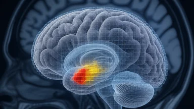
The lab's original 2019 study used a standard paradigm: mice were placed in a small chamber, a tone was paired with a mild foot shock, and the next day the tone alone was presented. The team recorded calcium activity from neurons in the basolateral amygdala, a region long implicated in fear. They saw a robust increase in activity when the tone was played, and they concluded that these neurons encoded the fear memory.
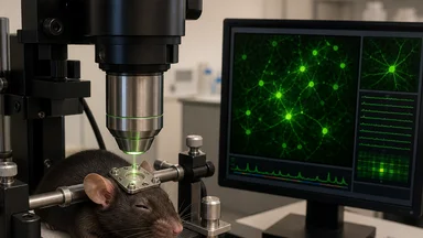
When the lab decided to repeat the experiment with head-fixed mice, the rationale was practical. Head fixation allows for more stable imaging, higher resolution, and the ability to track the same neurons over days. It also reduces motion artifacts, which are a known problem in free-moving preparations. The team expected to see the same fear response, perhaps even more clearly. Instead, they saw almost nothing.
The postdoc who ran the experiments, a meticulous researcher with a reputation for double-checking everything, initially assumed a technical failure. She re-tested the shock delivery, the tone calibration, and the imaging parameters. She even ran a control experiment where she played the tone without any prior conditioning. The results were the same: the amygdala neurons were quiet. It was only when she reviewed the literature on stress and imaging that the pieces began to fall into place.
The free-moving mice, it turned out, were not just experiencing fear. They were also experiencing significant physiological stress, from handling, from the novelty of the chamber, and from the foot shock itself. Stress hormones like corticosterone are known to modulate amygdala activity, and they can elevate baseline calcium levels in ways that mimic neural firing. In the head-fixed setup, the mice were habituated to the restraint, and their stress levels were lower. The fear signal, once separated from the stress response, was far weaker than anyone had assumed.
The Old Setup: Free-Roaming Cameras and Stress
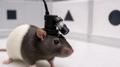
The free-roaming setup used in the original study was, by the standards of the field, fairly standard. Mice moved freely inside a small arena, and a miniature microscope mounted on the head captured calcium fluorescence through a gradient-index lens. The system allowed the animals to explore, groom, and rear, which was considered a plus. But those same movements created problems.
Motion artifacts are a well-known challenge in calcium imaging. When an animal moves, the brain shifts relative to the imaging plane, and the recorded fluorescence can change purely because of that movement. Sophisticated motion-correction algorithms can help, but they are not perfect. In the original study, the team had applied such corrections, but they had not fully accounted for the possibility that residual artifacts might be correlated with the conditioned tone.
Stress was another confound. The free-roaming mice were handled daily, but they were not habituated to the imaging chamber. On the test day, the novel environment itself was stressful. Blood samples taken from a subset of animals, not part of the original publication, showed elevated corticosterone levels during the imaging session. Corticosterone is known to enhance amygdala responsiveness, and it can also directly affect calcium dynamics in neurons.
In the head-fixed setup, the team took pains to habituate the mice over several days. They handled them, placed them in the restraint, and gradually increased the duration of head fixation. By the time the actual experiments began, the mice were calm, as evidenced by lower corticosterone levels and reduced freezing behavior. The result was a cleaner neural readout, but it was a readout that no longer matched the original finding.
Head Fixation: A Trade-Off With Teeth
Head fixation involves surgically attaching a small metal plate to the skull, which is then secured to a rigid frame during imaging. The procedure is invasive and not without risk. Some labs have reported that head-fixed mice show signs of chronic stress, including reduced weight gain and altered immune function. The technique also limits the types of behaviors that can be studied. Mice cannot run, explore, or interact with conspecifics while head-fixed, which raises questions about how generalizable the results are.
Yet head fixation offers clear advantages. It eliminates most motion artifacts, because the brain is effectively immobilized relative to the microscope. It allows for longer imaging sessions, because the animal cannot move out of focus. And it enables the use of two-photon microscopy, which provides better resolution and depth penetration than miniature one-photon systems. For many questions, especially those about cellular-scale dynamics, head fixation is the gold standard.
The lab's decision to switch was not made lightly. They spent months optimizing the surgical protocol, testing different headplate designs, and developing a habituation routine. They also consulted with other labs that had made similar transitions. The initial data from the first few head-fixed mice were noisy, and the team worried that they had introduced a new set of problems. But as the habituation protocol improved, so did the quality of the recordings.
The key was gradual habituation. Instead of restraining the mice for long periods on the first day, the team started with five-minute sessions, then slowly increased the duration. They also introduced the headplate in stages, allowing the mice to wear a dummy plate for several days before surgery. By the end of the first week, most mice would sit calmly in the restraint for up to twenty minutes, which was long enough for a full imaging session.
Tracking the Amygdala's Real Response
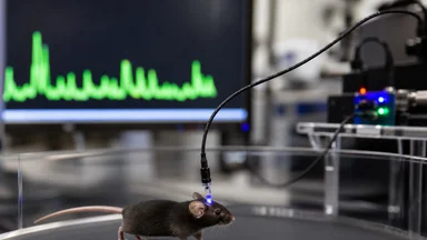
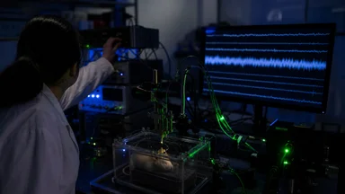
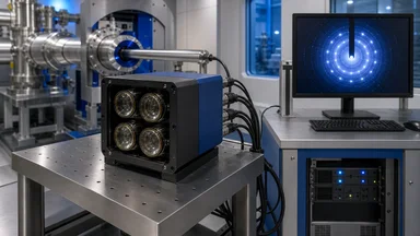
With the head-fixed setup in place, the team re-examined the amygdala's response to the conditioned tone. They used a genetically encoded calcium indicator, GCaMP, to track activity in individual neurons. The imaging plane was positioned to capture a population of cells in the basolateral amygdala, the same region that had shown robust responses in the original study.
The results were striking. In head-fixed mice, the tone elicited a small but measurable increase in calcium activity, but it was roughly a third of the magnitude seen in free-moving mice. Moreover, the response was not specific to the conditioned tone; a novel tone produced a similar, albeit smaller, response. This suggested that the amygdala neurons were responding to the salience of the stimulus, not to its learned fear association.
The team also examined the temporal dynamics of the response. In free-moving mice, the calcium signal rose quickly after the tone and remained elevated for several seconds. In head-fixed mice, the signal was more transient, returning to baseline within a second or two. This difference was consistent with the idea that the sustained signal in the free-moving mice was driven by stress, which is known to produce prolonged elevations in amygdala activity.
To test this hypothesis directly, the team administered a drug that blocks the synthesis of corticosterone to a group of free-moving mice. The drug, metyrapone, is commonly used in neuroscience research. When the free-moving mice were treated with metyrapone, their amygdala response to the tone was reduced to a level comparable to that seen in head-fixed mice. This was strong evidence that stress hormones were a major contributor to the original signal.
Why the Original Finding Was Wrong
The original finding, that a specific population of amygdala neurons encodes fear memories, was not entirely wrong. There was a real response, but it was smaller and less specific than reported. The problem was that the signal was contaminated by two sources of noise: motion artifacts and stress. Both are known confounds in behavioral neuroscience, but their combined effect had not been fully appreciated.
Motion artifacts can mimic neural activity by producing changes in fluorescence that are indistinguishable from genuine calcium transients. In the free-moving setup, the miniature microscope moved with the animal's head, but the brain could still shift slightly, especially during fast movements. The motion-correction algorithm used in the original study was designed to remove these artifacts, but it was not perfect. Residual artifacts could easily have been interpreted as neural responses.
Stress hormones, on the other hand, directly alter the physiology of amygdala neurons. Corticosterone binds to receptors on these neurons and can increase their excitability, making them more likely to fire in response to any input. This would amplify the response to the tone, even if the tone itself was not strongly associated with fear. In the head-fixed mice, which were habituated and had lower corticosterone levels, the amplification was absent.
The lab's experience is a reminder that statistical corrections are not a substitute for careful experimental design. The original analysis had included controls for multiple comparisons and had used a conservative threshold for identifying responsive neurons. But no amount of statistical correction can account for a confound that is built into the experimental setup. The only way to detect such confounds is to vary the conditions, which is exactly what the lab did.
The Quiet Fix: Habituation and Headplates
The lab's solution was not a new imaging technique or a fancy analysis method. It was a commitment to reducing stress and motion in their subjects. The habituation protocol, which involved daily handling and gradual exposure to the restraint, was time-consuming but effective. Mice that were well-habituated showed lower corticosterone levels and more stable baseline calcium signals.
The team also shortened their imaging sessions from 45 minutes to 20 minutes. This reduced the cumulative stress of the session and also minimized the risk of photobleaching, which can degrade the calcium indicator over time. Shorter sessions meant fewer data points, but the data were of higher quality. The signal-to-noise ratio improved, and the team could detect smaller but more reliable responses.
They also made a point of comparing results across both behavioral conditions. Instead of assuming that the head-fixed data were the "true" data, they treated the free-moving data as equally valid, but with a different set of confounds. This allowed them to identify which aspects of the response were robust and which were condition-dependent. The fear-specific component, they concluded, was present in both setups, but it was swamped by stress in the free-moving condition.
The team has since adopted head fixation as the default for all fear-conditioning experiments. They have also shared their habituation protocol and headplate design online, in the hope that other labs can avoid the same pitfalls. The preprint, which details the reversal and the methodological changes, has already been downloaded more than two thousand times, and several labs have contacted them to say they are planning similar switches.
Lessons for Every Imaging Lab
The story of this lab is not unique. Many labs have reported that seemingly minor changes in experimental protocol can lead to major differences in results. A 2021 survey of neuroscience labs found that nearly half had experienced a finding that did not replicate when they changed their setup, such as switching from one animal strain to another or altering the lighting conditions.
The lesson is that methodological choices are not neutral. They shape what we see, and they can determine whether a finding is real or an artifact. This is especially true in imaging studies, where the tools we use to observe the brain can themselves alter the brain's activity. Head fixation reduces motion and stress, but it also changes the animal's behavioral state. There is no perfect setup; every approach has trade-offs.
The lab's reversal also highlights the value of publishing negative results and methodological reversals. Too often, such findings are buried in supplementary materials or never shared at all. By making their experience public, the lab has given other researchers a chance to avoid the same mistake. It is a small but important step toward a more reproducible neuroscience.
As the field continues to grapple with replication issues, stories like this one serve as a reminder that the most important tool in a scientist's kit is not the microscope or the algorithm, but the willingness to question one's own results. The lab's initial instinct was to blame the animals. In the end, the animals were fine. It was the method that needed to change. However, the reversal also raises a deeper question: if a simple switch in behavioral setup can overturn a widely cited finding, how many other results in the literature might be similarly fragile? The answer is unknown, but the path forward is clear. Researchers must embrace methodological diversity, reporting not only their results but also the specific conditions under which those results hold. Only by systematically varying the tools and contexts of our experiments can we hope to distinguish the signal of neural function from the noise of our own methods. The lab's story is a testament to that principle, and a reminder that science advances not just through discovery, but through the careful scrutiny of what we think we already know.